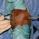
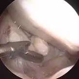
Arthroscopy is a minimally invasive joint surgery procedure that can be performed for diagnostic or therapeutic purposes. This keyhole surgical technique enables to visualise the joint surface using a special camera (arthroscope) after filling the joint with sterile saline solution or gas. Surgical procedures can then be carried out in the joint through a second instrument portal. These portals are usually less than one centimetre in size. This minimises trauma to the joint and speeds up the horse's recovery.
Arthroscopic operations are one of our clinic's main areas of specialisation. As one of the first clinics to perform arthroscopies in Germany, we have decades of experience in this field.
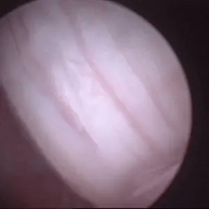
Diagnostic arthroscopy can provide information about the cause, treatment options and prognosis of the disease in the case of unexplained lameness that has been localised to a specific joint through a thorough orthopaedic examination. As part of this examination, the cartilage surface, tendon and ligament structures or the menisci can be examined.
In addition to its diagnostic benefits, arthroscopy is above all an efficient and gentle treatment option for various diseases of the joints, tendon sheaths and bursae.
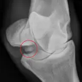
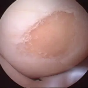
The most common indication for arthroscopy is the removal of osteochondral fragments (osteochondrosis dissecans / OCD / chip) or splinters from the joint and the smoothing of cartilage damage that can occur as a result. These so-called “chip surgeries” are performed when clinical complaints such as joint effusion and/or lameness occur due to a chip. Due to the high requirements associated with the marketing of sport horses nowadays, many horses will also undergo chip surgery prior to sale. The aim of this is to improve marketing opportunities on the one hand and to avoid possible joint damage caused by friction of the chips in the joint on the other, thus ensuring that the athlete remains healthy in the long term.
Chips are most frequently found and operated on in the hoof, fetlock, hock and stifle joints. However, we also offer arthroscopic removal of bone fragments in the pastern, carpal (knee), elbow and shoulder joints.
In addition to the removal of chips and fragments, we can perform surgical cyst treatments in the various joints arthroscopically, depending on their location and size.
In the area of the stifle, surgical treatment of meniscal damage is also possible at our clinic. This involves the surgical removal of partial tears of the meniscus. For infected joints, arthroscopy offers the best option for joint irrigation, as the high irrigation pressure and the use of large cannulas allows to flush out bacteria and inflammatory products more effectively, which significantly improves the survival rate compared to conservative treatment. It is also possible to remove foreign bodies from the joint under visual control. In adult horses, infected joints usually develop because of a traumatic injury with traumatic tearing of the joint capsule, while in foals pathogens can also enter the joint via the blood (haematogenous). In addition to joint diseases, diseases of the tendon sheaths, in particular the digital flexor tendon sheath, are also an indication for endoscopic surgery. Here we offer surgical treatment of tendon fraying and adhesions as well as desmotomy of the annular ligament and resection of the manica flexoria. Diagnostic endoscopy is also possible here. Endoscopy of the carpal tendon sheath may be indicated in the case of carpal tunnel syndrome or for the removal of so-called osteochondromas. In foals or young horses with flexural deformities or in adult horses with tendon damage, the supporting ligament of the superficial flexor tendon can be cut endoscopically in a minimally invasive procedure. In diseases of the podotrochlear apparatus, such as damage to the deep digital flexor tendon inside the hoof capsule an endoscopic examination (bursoscopy) of the podotrochlear bursa with debridement of the defect can improve the chances of recovery. Should an infection of this bursa occur, for example as a result of a street nail injury, bursoscopy can also significantly improve the prognosis. The same applies to infections of other bursae, for example in the ankle (bursa subcutanea/bursa subtendinea) or in the shoulder (bicipital bursa).
For infected joints, arthroscopy offers the best option for joint irrigation, as the high irrigation pressure and corresponding irrigation cannulas allow bacteria and inflammatory products to be flushed out more effectively, which significantly improves the survival rate compared to conservative treatment. It is also possible to remove foreign bodies from the joint under visual control. In adult horses, infected joints usually develop as a result of a traumatic injury with opening of the joint capsule, while in foals pathogens can also enter the joint via the blood (haematogenous) as part of the foal lameness complex.
In addition to joint diseases, diseases of the tendon sheaths, in particular the so-called fetlock flexor tendon sheath, are also an indication for endoscopic surgery. Here we offer surgical treatment of tendon fraying and adhesions as well as cutting of the fetlock ring ligament and resection of the manica flexoria. Diagnostic endoscopy is also possible here. Endoscopy of the carpal tendon sheath may be indicated in the case of carpal tunnel syndrome or for the removal of so-called osteochondromas. In foals or young horses with a tendon foot or in adult horses with tendon damage, the supporting ligament of the superficial flexor tendon can be cut endoscopically in a minimally invasive procedure.
In the context of hoof roll diseases, such as damage to the deep flexor tendon in the area of the hoof roll, an endoscopy (bursoscopy) of the hoof roll bursa with smoothing of the defect can improve the chances of recovery. Should an infection of this bursa occur, for example as a result of a nail bite, bursoscopy can also significantly improve the prognosis. The same applies to infections of other bursae, for example in the ankle (bursa subcutanea/bursa subtendinea) or in the shoulder (bicipital bursa).