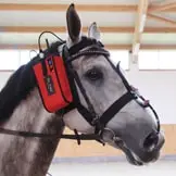
Endoscopy allows the examination of body cavities and hollow organs, facilitating the diagnosis of diseases and, in some cases, immediate therapeutic intervention. In veterinary medicine, endoscopic examinations are primarily used to assess the upper airways (pharynx, larynx, and guttural pouches) as well as the lower airways. The stomach can also be evaluated using a 3-metre-long endoscope (gastroscopy). Additional organs that may be examined include the uterus and the urinary bladder, for example in cases of haematuria.
At our clinic, flexible video endoscopes are used, enabling the recording and storage of high-quality images and video sequences. A rigid video endoscope is employed for dental examinations. Various instruments can be introduced through the endoscope’s working channel. For example, biopsy forceps allow the collection of tissue samples for cytological or histopathological examination, and a high-frequency laser enables minor surgical procedures such as incision or removal of cysts or tumours under endoscopic guidance. Medications can also be administered with high precision.
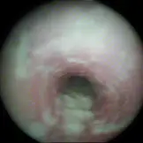
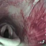
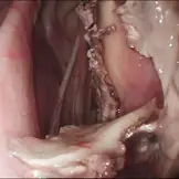
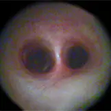
Laryngoscopy
Laryngoscopy is the endoscopic examination of the pharynx and larynx. It is most commonly indicated in horses presenting with inspiratory or expiratory respiratory noise during exercise, such as in cases of laryngeal hemiplegia or dorsal displacement of the soft palate. Upon request, laryngoscopy can also be performed dynamically while the horse is being ridden or lunged.
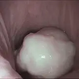
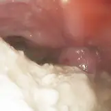
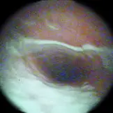
Guttural Pouch Endoscopy
Horses possess unique anatomical structures known as guttural pouches, which are air-filled diverticula of the Eustachian tubes located ventral to the base of the skull. Because the guttural pouches communicate with the nasopharynx via the auditory tube, infectious agents can readily enter this space. Indications for guttural pouch endoscopy include enlargement of the retropharyngeal lymph nodes as well as unilateral bloody or purulent nasal discharge.
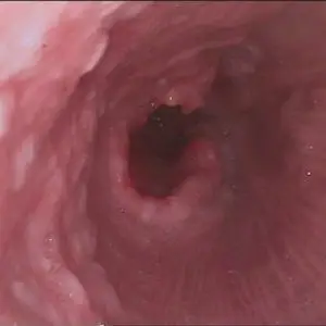
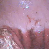
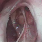
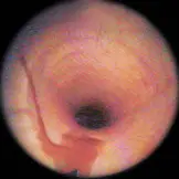
Bronchoscopy
Every comprehensive airway examination should include bronchoscopy. The endoscope is advanced through the ventral nasal meatus into the pharynx. The trachea is entered via the laryngeal opening and can be followed to its bifurcation into the main bronchi. If tracheobronchial secretions are present, they can be collected directly using a sampling catheter passed through the working channel and subsequently analysed.
If only small amounts of secretion are present, a bronchoalveolar lavage (BAL) can be performed. In this procedure, a sterile fluid is instilled into the lower airways and subsequently aspirated for laboratory analysis.
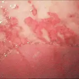
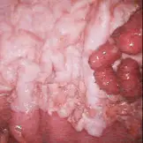
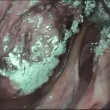
Gastroscopy
Gastroscopy allows visualisation of the oesophagus, stomach, and the proximal portion of the small intestine (duodenum). Prior to the procedure, the horse should be fasted for at least 15 hours, with water withheld for approximately four hours. This ensures that the stomach is physiologically empty, allowing thorough evaluation of the gastric mucosa.