Magnetic resonance imaging (MRI) is one of the most modern imaging procedures in equine medicine. It works entirely without X-rays or radioactive substances and is based on a combination of magnetic fields and radio waves.
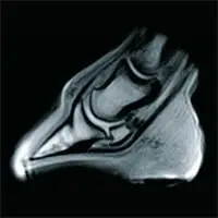
The MRI examination enables a particularly detailed visualisation of soft tissue structures and bones, especially in the limbs. It is particularly valuable for diagnostics in the hoof area, as this is where important structures such as the deep flexor tendon or bursa are located, which are difficult to visualise using other imaging techniques.
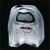
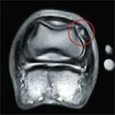
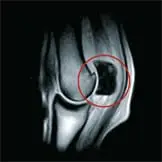
Changes in the bone marrow, known as bone oedema, can also be visualised. These accumulations of fluid in the bone are caused, for example, by overloading, microfractures or inflammation. If they are recognised in good time, the therapy can be adapted in a targeted manner to prevent more serious damage such as stress fractures or chronic lameness.
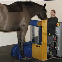
With our Hallmarq low-field magnet (0.27 Tesla), regions from the hoof to the carpal joint on the front leg or to the hock joint on the hind leg can be examined on a standing horse.