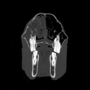
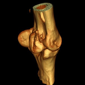
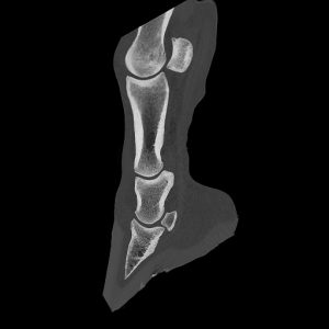
Computed tomography, like magnetic resonance imaging, is a cross-sectional imaging procedure. Unlike MRI, CT images are produced with the help of X-rays. The horse is x-rayed layer by layer and a computer calculates a three-dimensional image of the examined region from the numerous individual images. This makes it possible to visualise even very small changes in bones or soft tissue that would not be visible with conventional X-ray images. If necessary, a contrast agent can also be used to visualise certain structures even better.
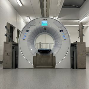
Our powerful CT scanner (Qualibra Exeed, equipped with Aquilion Exceed from Canon Medical and artificial intelligence from AiCE) with a particularly large opening (gantry) of 90cm enables, among other things, the visualisation of previously inaccessible body regions such as the pelvis, shoulder region and other parts of the spine.
A particular advantage of our system is the ability to perform many examinations on a standing, sedated horse. Thanks to a pit in which the CT device can not only move forwards and backwards, but also up and down, the head, parts of the cervical spine and the lower limbs (up to the tarsal or hock joint) can be examined without general anaesthesia. This reduces the risk for the horse and enables a gentler procedure.
Computed tomography examinations of the upper limb and posterior sections of the cervical spine require a general anaesthetic, in which the horses are placed under general anaesthetic as for an operation and can then be driven through the operating theatre into the directly adjacent CT room. This allows the general anaesthetic to run smoothly and also enables operations to be performed under CT control.